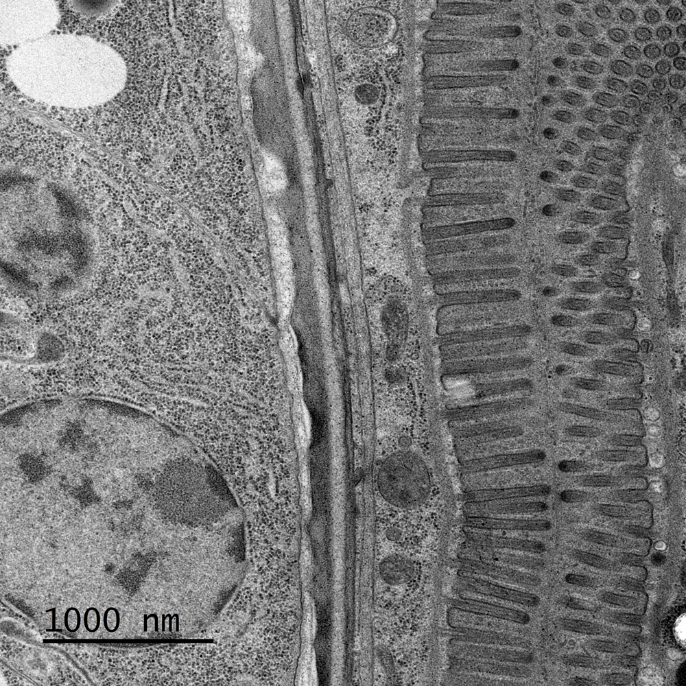
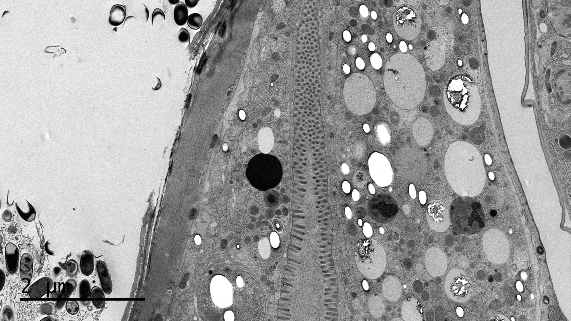
High-resolution image of C. elegans stomach sample
Captured with the Rio 16 camera; 200 kV; TEM indicated magnification: 6.3kx; image size: 4k x 4k; exposure time: 1 s.
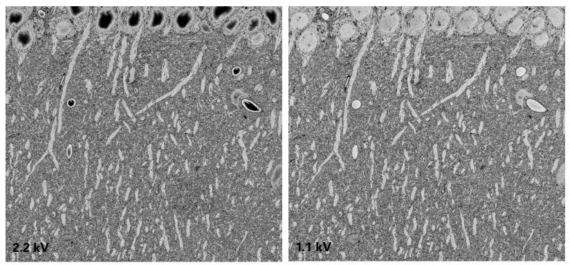
OnPoint detector minimizes charging and beam damage
Image courtesy of T. Deerinck NCMIR/UCSD.
Image shows that the OnPoint BSE detector can resolve features at 1.1 kV (right) that were previously distorted (e.g., black spots) by charging or beam damage at 2.2 kV (left). Sample: Mouse cerebellum.
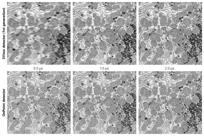
OnPoint increases 3View imaging speed four-fold
Compares image quality from the 1st generation 3View® detector (top) to the OnPoint™ detector (bottom) at various dwell times. Results show the OnPoint detector delivers equal or better resolution images using shorter dwell times (0.5 vs. 2.0 µs). Sample: Brain, 3 kV, 15 kx, 0.5 µs dwell time.
Preserves synaptic vesicle under low kV conditions
Image courtesy of T. Deerinck NCMIR/UCSD.
Demonstrates how low kV conditions can preserve and discriminate fine features, such as synaptic vesicles. Sample: 2 kV, 0.2 nm pixels, 24k x 24k image, 1 µs dwell time.
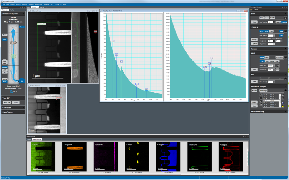
Gatan Microscopy Suite Software
EELS made easy with the new Gatan Microscopy Suite® (GMS) 3 software.
- Advanced model-based quantification and mapping
- STEM SI acquisition modalities
- Experiment oriented workflow for fast time to results
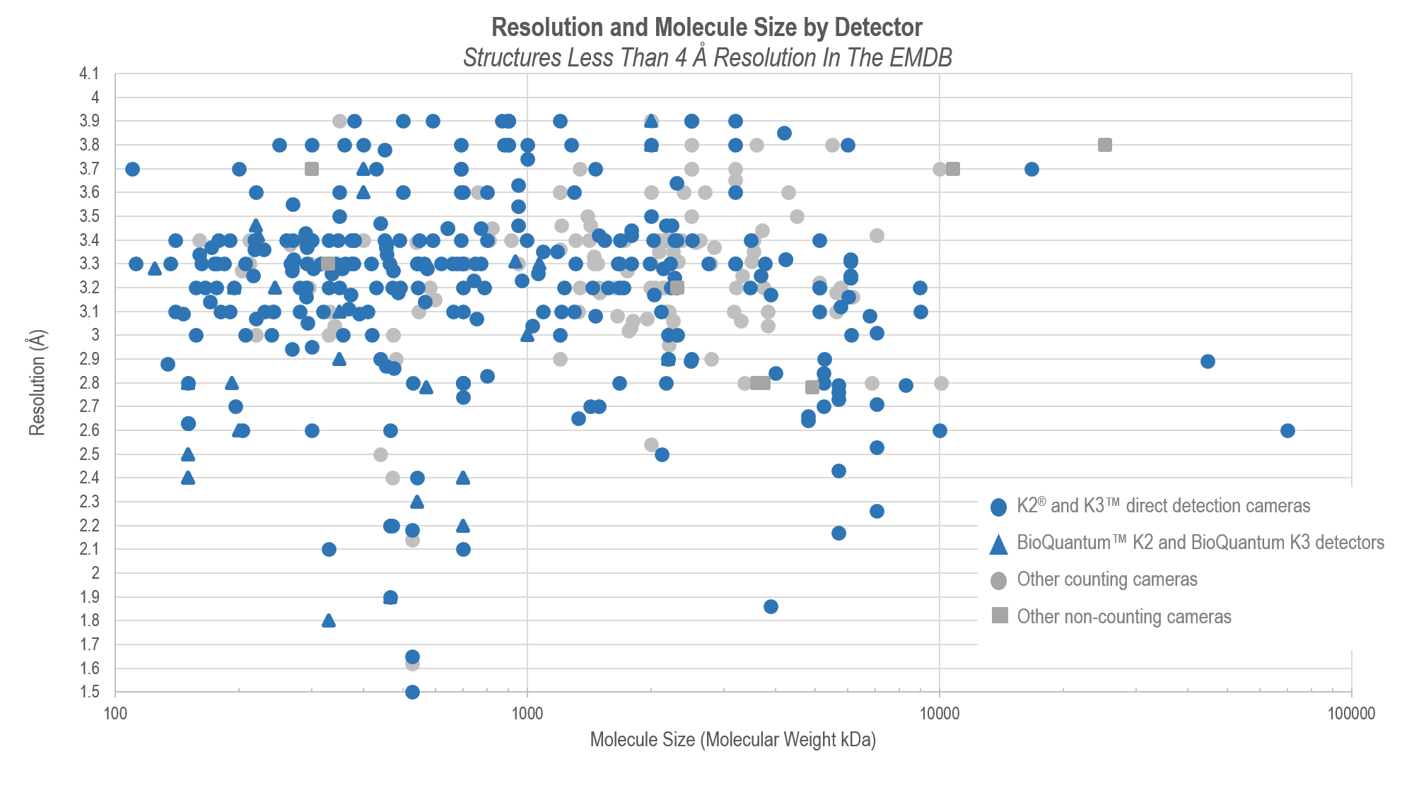
The Efficient Frontier of Resolution
Comparison between the resolution and molecule size for published single-particle cryo-EM structures.
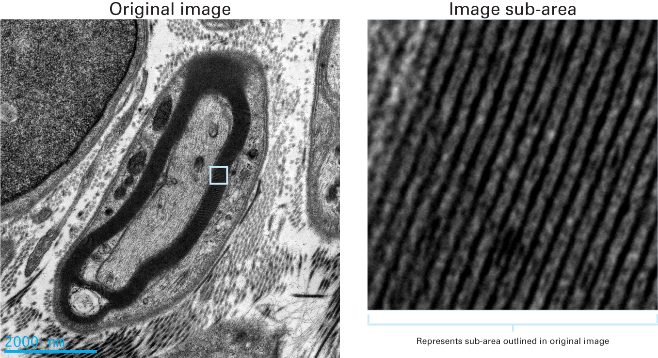
Clearly resolved myelin sheaths and collagen fibers
The zoomed in image (right) obtained using the OneView camera shows clearly-defined detail within the myelin sheathes and collagen fibers when compared to the original image (left).
Sample: skin; beam energy: 120 kV; image size: 4k x 4k; exposure: 1 s; number of frames: 25
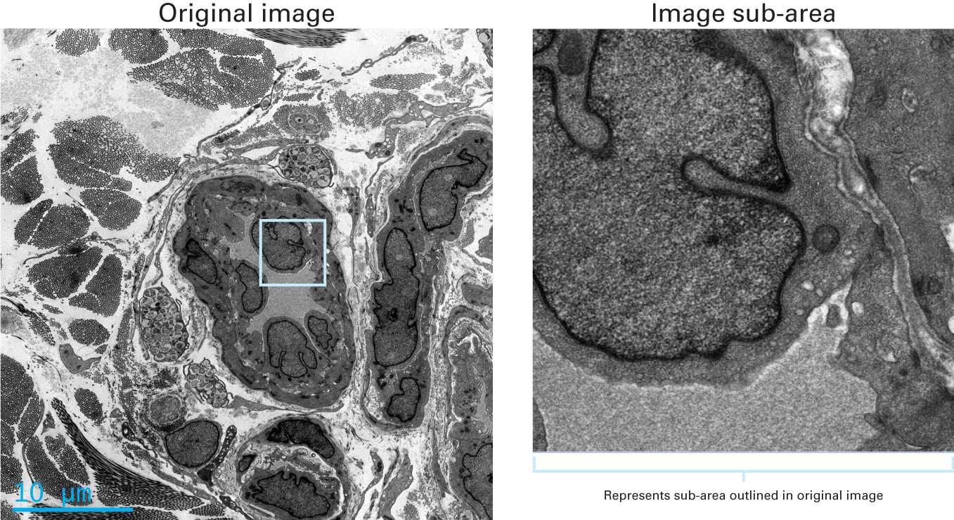
Fine details revealed in skin cells
The zoomed in image (right) obtained using the OneView camera shows clearly-defined detail and resolution when compared to the original image (left).
Sample: skin; beam energy: 120 kV; image size: 4k x 4k; exposure: 1 s; number of frames: 25
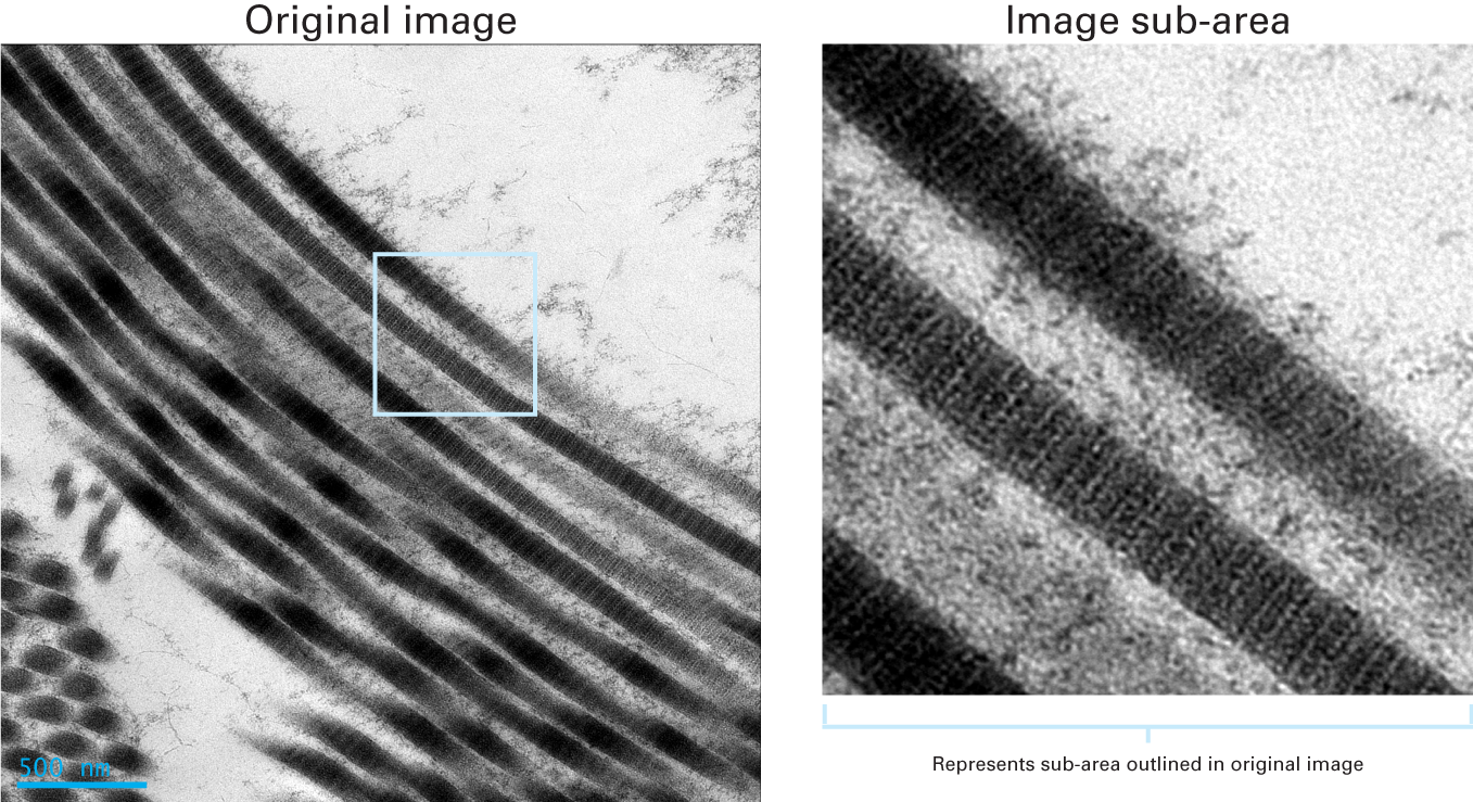
Clearly resolved collagen fibrils
Collagen fibrils are clearly resolved in this 4k x 4k image obtained using the OneView camera. This definition is maintained when the original image is zoomed in on during post-processing.
Sample: collagen; beam energy: 120 kV; image size: 4k x 4k; exposure: 1 s; number of frames: 25
High resolution image of C. elegans
OneView camera captures high resolution images of biological specimens, such as C.elegans at 3840 x 2160 pixels.
Pages
