Paolo Longo, Ph.D., Gatan Inc.
Sample courtesy of Dr. Joshua Sugar at Sandia National Lab, Livermore, CA
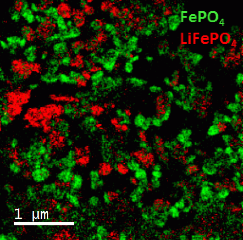
EELS color map showing the distribution LiFePO4 (red) and FePO4 (green) particles from a battery electrode charged to half cycle
Methods
FEI F20 TEM/STEM microscope; S-FEG emission gun; GIF Quantum® ER system; voltage: 200 kV; EFTEM SI mode from 680 – 730 eV; slit width: 2 eV ; step size: 1 eV; total exposure time: <4 min

